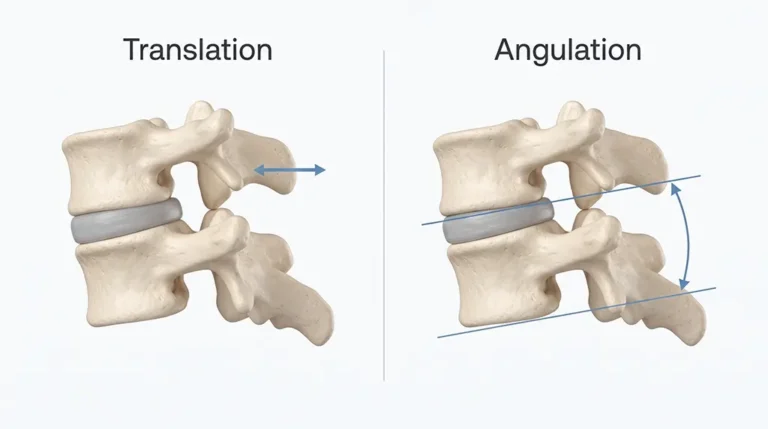
If you are reviewing a Digital Motion X-Ray report after a cervical injury, two terms may appear repeatedly: translation and angulation.
For patients, healthcare professionals, and attorneys, understanding these terms is important because they describe how one cervical vertebra moves relative to another. These measurements help describe the functional biomechanics of the cervical spine.
Digital Motion X-Ray at DMX Miami evaluates these relationships while the cervical spine is upright, weight-bearing, and moving.
DMX Miami works with patients and referring professionals from Miami, Fort Lauderdale, Miami-Dade County, Broward County, the Florida Keys, other parts of Florida and the United States, as well as international patients from Colombia, Chile, Argentina, Mexico, and the Caribbean.
What Is Vertebral Translation?
Translation is linear movement of one vertebra relative to an adjacent vertebra.
In simple terms, one vertebra slides relative to another. Some translation is normal. A healthy cervical spine must move. The clinical question is whether that movement remains appropriately controlled.
The examiner may evaluate how much translation occurs, when it occurs, its direction, whether one level behaves differently from neighboring levels, and whether the pattern correlates with the patient’s symptoms and injury.
What Is Angulation?
Angulation is the change in angle between adjacent vertebral segments.
When the neck flexes or extends, the vertebrae normally change their angular relationship. The clinical question is whether the amount of angulation is appropriate, whether the motion occurs smoothly, and whether one segment contributes excessively compared with neighboring levels.
Why Aren’t Translation and Angulation Automatically Abnormal?
Because normal joints move. The goal is not a spine with zero motion. The goal is controlled motion.
A properly functioning motion segment must allow enough movement for normal function while preventing excessive movement that could compromise stability. That balance depends heavily on the stabilizing structures.
What Role Do Ligaments Play?
Ligaments are mechanical restraints. They connect bone to bone and help control how much one vertebra can move relative to another. Their functions include limiting excessive translation and angulation, controlling rotation, guiding normal movement, and maintaining joint stability.
Therefore, the functional integrity of a ligament is directly related to the movement it is designed to control. For more background on how trauma can affect this restraint system, see Whiplash Injuries and Cervical Instability.
Why Must the Joint Move to Evaluate Ligament Function?
Consider what a ligament actually does. If its job is to prevent excessive movement, its mechanical performance cannot be fully evaluated by observing only a joint that is not moving.
Static imaging provides information about structure. But to evaluate the mechanical restraint provided by the ligamentous system, the joint must move.
“When the cervical spine moves under load, do the ligaments adequately restrain the vertebral segments?”
For a patient-focused explanation of this concept, read Why Do My Neck Symptoms Happen Only When I Move?.
Why Does Weight Bearing Matter?
The cervical spine normally functions while supporting the weight of the head. DMX evaluates the neck upright, under gravity, weight-bearing, and during controlled movement.
Those conditions place the stabilizing system under functional demand. The ligaments and muscles must control motion while the head is being supported. This allows the examination to evaluate function rather than appearance alone.
How Can Ligament Injury Affect Translation?
If ligamentous restraint is reduced, one possible consequence is excessive vertebral translation. The motion segment may slide farther than expected during movement.
The clinical significance depends on magnitude, spinal level, direction, symptom correlation, injury history, and associated findings.
How Can Ligament Injury Affect Angulation?
Compromised ligamentous restraint may also permit excessive angular movement between adjacent vertebrae. The pattern must again be evaluated in clinical context.
What Is Motion Sequencing?
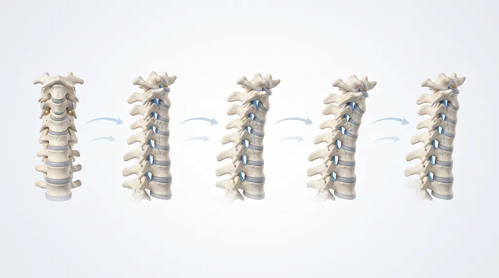
The cervical spine does not move as one rigid unit. Movement is distributed through several segments. Motion sequencing describes when and how each segment contributes to the movement.
Dynamic imaging may identify patterns such as one level moving unusually early, one segment moving excessively, another segment remaining relatively restricted, abrupt changes in movement, or directional asymmetry.
This provides information that cannot be obtained simply by comparing the beginning and ending positions.
How Is This Different From Flexion-Extension X-Ray?
Traditional flexion-extension radiographs generally provide selected still images in flexion and extension. They can provide valuable information about endpoint positions. DMX records the movement sequence itself.
The distinction is similar to photographs versus video. Endpoint images show where the vertebrae ended. A motion study also shows how they got there.
For another comparison of static and dynamic approaches, see Digital Motion X-Ray vs. Upright MRI.
How Do MRI, CT, X-Ray, and DMX Fit Together?
| Study | Primary Role |
|---|---|
| CT | Detailed bony anatomy and many acute traumatic injuries |
| MRI | Spinal cord, discs, nerve roots, and many soft tissues |
| Static X-Ray | Alignment and bony relationships in selected positions |
| DMX | Upright, weight-bearing function; intersegmental translation, angulation, symmetry, sequencing, and ligament-controlled stability |
These studies provide different categories of information.
Why Is Clinical Correlation Important?
Motion measurements should be interpreted in the context of the patient. Relevant information includes accident mechanism, onset of symptoms, previous injuries, degenerative findings, examination, neurological findings, other imaging, and movement-triggered symptoms.
Two patients can have similar measurements but very different clinical circumstances. The question is not simply “Is there movement?” The question is:
“Is this movement biomechanically abnormal and clinically relevant for this patient?”
What Should Referring Providers Look for in a DMX Report?
A useful dynamic report should identify which movements were tested, which spinal levels were evaluated, translation measurements, angular measurements, relevant asymmetries, upper-cervical findings when applicable, measurement methodology, and other radiographic findings.
You can also review a sample DMX report from DMX Miami.
What Should Attorneys Understand About DMX Measurements?
For injury cases, translation and angulation can provide objective measurements of cervical motion. The medical significance of those measurements should be considered together with the mechanism of injury, medical history, symptom onset, examination, previous imaging, and treatment records.
Dynamic imaging can contribute objective biomechanical documentation. Broader medical and legal conclusions require the complete clinical record.
For the patient-facing whiplash pathway, see Whiplash Imaging in Miami.
Professional Referrals to DMX Miami
DMX Miami works with chiropractors, physicians, physical therapists, rehabilitation professionals, regenerative-medicine providers, and personal-injury attorneys and their provider networks.
Our goal is to provide clear, objective information about upright, weight-bearing cervical motion and functional biomechanics.
For patient evaluations or professional referrals, contact DMX Miami or call 305-275-7474.
Frequently Asked Questions
What is translation on a DMX report?
Translation is linear sliding of one vertebra relative to an adjacent vertebra.
What is angulation?
Angulation is the change in angle between adjacent vertebral segments during movement.
Is all translation abnormal?
No. Normal cervical joints move. The amount, pattern, and control of movement determine its clinical significance.
How do ligaments affect translation and angulation?
Ligaments mechanically restrain excessive vertebral movement. Compromised ligamentous restraint can therefore lead to abnormal translation or angulation.
Why is DMX performed weight-bearing?
The cervical spine normally supports the head under gravity. Upright imaging evaluates the stabilizing system while it is functioning under that load.
Why does the joint have to move to evaluate ligament function?
Because the mechanical role of a ligament is to control movement. Functional ligament integrity is evaluated by determining whether the joint remains appropriately restrained during motion.
Can DMX and MRI both be useful?
Yes. MRI primarily evaluates structural anatomy, while DMX provides information about functional biomechanics during weight-bearing movement.
References
- Wang et al. – The Quantitative Measurements of the Intervertebral Angulation and Translation During Cervical Flexion and Extension
- Published cervical-motion and videofluoroscopy literature
- American College of Radiology / RadiologyInfo – Suspected Spine Trauma
Footer: DMX Miami serves patients in Miami, Fort Lauderdale, Miami-Dade, Broward, the Florida Keys, Florida, the USA, and visitors from Colombia, Chile, Argentina, Mexico, and the Caribbean.
Learn more: Treatment
Schedule your appointment today:Appointments
Call 305-275-7475 orbook your appointment online

Dr. Rodolfo Alfonso, D.C.
Dr. Mark N. Berry, D.C.
Sunset Chiropractic and Wellness
8585 Sunset Dr. STE 102
Miami, Florida 33143