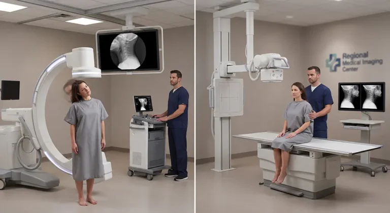
Flexion-extension X-rays and Digital Motion X-Ray (DMX) both evaluate the spine beyond a single neutral image, but they do so differently. Traditional flexion-extension radiographs usually capture still images near the end points of flexion and extension. DMX uses fluoroscopic X-ray technology to record movement continuously as the patient moves through a controlled range. That can provide information about when motion occurs, how vertebral segments move relative to one another, and whether motion appears asymmetric or concentrated at particular levels. Neither examination should be interpreted in isolation, and more motion data does not automatically mean a clinically important abnormality is present.
- Flexion-extension X-rays typically compare selected static positions.
- DMX records continuous spinal movement.
- End-position measurements and motion sequencing answer related but different questions.
- Neither test should replace the patient’s history, examination, MRI, CT or neurological assessment when those are indicated.
- Dynamic findings must be interpreted in clinical context.
Last updated: September 29, 2026
Reviewed by: DMX Miami clinical team
Patients frequently arrive at DMX Miami and say:
“I already had flexion-extension X-rays. Isn’t that the same thing?”
They are related.
But they are not identical.
Understanding the difference begins with understanding what each examination records.
What Are Flexion-Extension X-Rays?
Traditional flexion-extension radiographs usually involve taking separate X-ray images while the patient bends the neck or low back:
- forward
- backward
The provider can then compare alignment between those positions.
Depending on the examination and region, measurements may be made involving:
- vertebral translation
- angular relationships
- alignment
- range between positions
This can be useful information.
But the result generally consists of individual images representing selected points in the patient’s range of motion.
What Is Digital Motion X-Ray?
Digital Motion X-Ray uses fluoroscopic X-ray imaging to record the spine while movement is actually occurring.
Instead of seeing only:
Position A → Position B
the examiner can observe what happens between those positions.
Conceptually, the difference is similar to:
photographs versus video.
Both can provide useful information.
They simply present movement differently.
Why the Middle of the Motion Can Matter
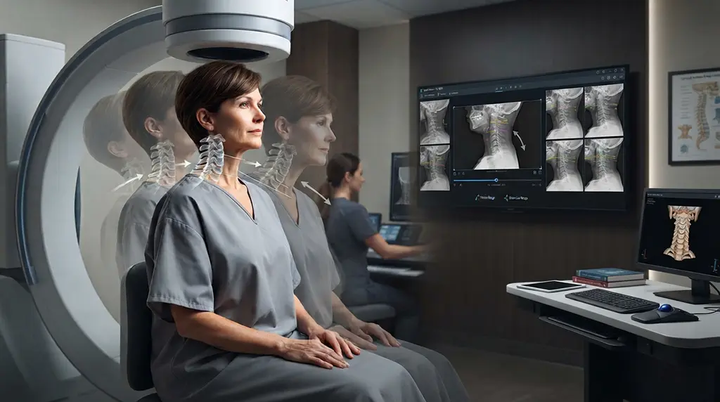
Imagine that five cervical vertebral segments contribute to flexion.
In an ideal movement pattern, motion is distributed in an organized way.
But suppose:
- Several segments move very little initially.
- One segment moves disproportionately.
- Another segment catches up later.
- The final flexed position looks relatively acceptable.
Two endpoint images might provide useful measurements of the starting and ending positions.
Continuous imaging can additionally show the sequence used to get there.
That does not automatically mean the sequence is pathological.
But it provides another category of information.
Translation
Translation refers to sliding motion between vertebral segments.
Providers evaluating cervical or lumbar mechanics may consider how much translation occurs and when it occurs during movement.
Angulation
Angulation describes changes in the angular relationship between adjacent vertebrae.
Again, these measurements should be interpreted with appropriate standards and the patient’s overall clinical picture.
Motion Sequencing
DMX can also allow observation of whether different spinal levels appear to participate smoothly throughout movement.
Potential observations may include:
- delayed movement
- abrupt movement
- asymmetry
- disproportionate motion at one level
- hinge-like behavior
These observations require careful interpretation.
Not every unusual movement pattern represents ligament instability or injury.
Why Flexion-Extension X-Rays Are Still Useful
DMX should not be marketed by suggesting that ordinary flexion-extension radiographs are useless.
They are not.
Flexion-extension films can provide valuable information about:
- spinal alignment
- positional translation
- positional angulation
- range between selected positions
The correct question is:
What clinical information are we trying to obtain?
When Continuous Motion May Be Particularly Interesting
Consider a patient who reports:
- pain only midway through rotation
- a cervical catch during extension
- clicking during movement
- symptoms returning from flexion
- one direction feeling different from the other
- headaches triggered at a specific point in motion
In such a patient, observing the movement sequence may provide information that endpoint positions alone cannot provide.
DMX Does Not Replace MRI or CT
This remains an important part of our content strategy.
MRI may provide valuable information about:
- discs
- spinal cord
- nerve structures
- soft tissues
- other anatomical pathology
CT may provide detailed information about:
- bone
- fractures
- traumatic anatomy
DMX asks another question:
How do the vertebrae behave during motion?
A complete evaluation may involve more than one imaging modality.
Why Symptoms Still Matter
An imaging finding without symptoms or functional relevance should not automatically drive treatment.
Likewise, symptoms cannot be dismissed just because one imaging examination appears relatively normal.
The provider must combine:
- accident mechanism
- symptoms
- examination
- neurological findings
- prior imaging
- functional limitations
- dynamic findings when appropriate
How Findings May Change Care
When clinically significant abnormal movement is identified, care may emphasize:
- cervical or lumbar stabilization
- controlled motion retraining
- improved endurance
- avoidance of repeatedly provocative positions
- modification of manual treatment
- functional rehabilitation
- coordination with other providers
The purpose of imaging is to improve clinical decision-making.
FAQs
Is DMX the same as flexion-extension X-ray?
No. Both evaluate the spine in more than one position, but DMX records continuous fluoroscopic movement while traditional flexion-extension studies generally use selected still images.
Does DMX automatically find problems that flexion-extension X-rays miss?
No. Each examination provides different information, and neither guarantees that a clinically significant abnormality will be identified.
Is DMX better than MRI?
They answer different questions. MRI provides detailed static anatomical information, while DMX evaluates movement.
Can DMX diagnose ligament instability by itself?
No. Dynamic findings should be interpreted alongside symptoms, examination, history and other imaging.
References
- Cleveland Clinic: Whiplash
- Published literature on cervical videofluoroscopy and dynamic spinal assessment
- Published literature on flexion-extension radiography and segmental spinal motion
Footer: DMX Miami serves patients in Miami, Fort Lauderdale, Miami-Dade, Broward, the Florida Keys, Florida, the USA, and visitors from Colombia, Chile, Argentina, Mexico, and the Caribbean.
Learn more: Treatment
Schedule your appointment today:Appointments
Call 305-275-7475 orbook your appointment online

Dr. Rodolfo Alfonso, D.C.
Dr. Mark N. Berry, D.C.
Sunset Chiropractic and Wellness
8585 Sunset Dr. STE 102
Miami, Florida 33143